
 | ||
| Paua shell overlaid onto SEM image. |
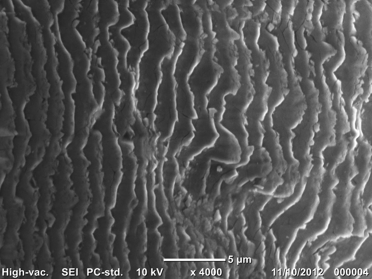
The Photon Factory, my workplace, just got a new table-top scanning electron microscope (SEM), so I thought I would take some images of a Paua shell under the microscope.

The Paua shell is New Zealand's national shell. However, few know why it has opalescent colouring. This is due to how the mollusc builds its shell using layers of microscopic calcium carbonate tiles stacked like bricks. (Here is a 3D structure of the argonite)
 |
| Paua shell we had in the lab |

What else would you like to see under the new SEM?Using GIMP I overlaid the picture with the the colours of the Paua shell.