Showing posts with label Raman spectroscopy. Show all posts

Tuesday, 14 February 2017

Chemical analysis of milk on a compact disc

We know how much food colouring has been added to a recipe by the intensity of the colour. Food colouring is made of dye molecules which absorb certain colours of light and scatter others. Measuring the concentration of food colouring is easy - all that is needed is a light emitting diode (LED) and a light sensor (link to laser cut detector). But how do we determine the concentration of molecules that are not coloured? One method that has recently gained a lot of attention is Raman spectroscopy. Put simply, a laser is used to excite the molecules, then a camera picks up a characteristic fingerprint derived from the way the molecules jiggle/vibrate due to thermal motion.

Michel Nieuwoudt and others in the Photon Factory have found that Raman spectroscopy can allow for the determination of all of the important components in milk such as protein, fat and health indicators. In a previous post I wrote about measuring the contaminant melamine in milk using gold coated Blu-ray discs which amplified the weak Raman signal to something detectable. This month we published a follow-up work that aimed to integrate milk analysis into a device that could be used in a dairy shed.
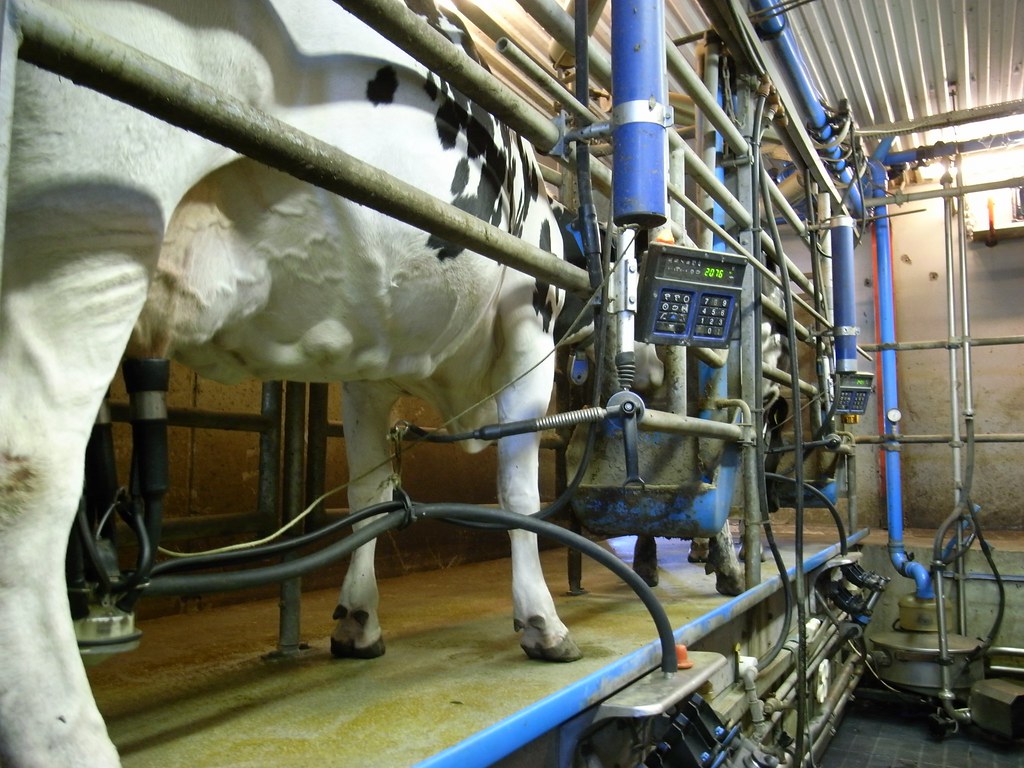
Giving milk
CC BY-NC 2.0 Giving Milk by Morton Just

The first challenge was finding a device into which to integrate the analysis. We wanted to make use of microfluidic technology, which uses techniques from the semiconductor industry to make very small fluid channels on the micron scale (your hair is about 100 microns across). Advantages include being able to pack many tests onto a single device and having very precise control over the liquid. Traditional microfluidic requires a lab full of pumps to operate, which is not conducive for analysis in a dairy shed, so we turned to the newer field of centrifugal microfluidics. Combining compact disc technology and microfluidics allows for standalone operation with pumping driven by the centrifugal force as the liquid pipetted into the centre of the disc is spun outwards through microfluidic channels.

CC BY 3.0 LabDisk for SAXS

To make this approach viable we have to use injection-moulded plastic discs to keep the costs down and to allow for large scale production of many discs that can be delivered to the farmer.

The problem with performing Raman analysis in a plastic device is that the plastic has a very strong Raman signal, which drowns out the weak Raman signal from the milk. In order to solve this problem we removed the plastic between the laser and the milk - holding the milk in an open channel using the capillary force. This is the same force that pulls water up the sides of a glass to form a meniscus.

Cross sections of the multilayer disc with the laser cut channel on the bottom, a layer of double sided tape in the middle and a top cover which leaves some of the channel open

The main contribution of the paper was working out how to use the centrifugal force to fill the channel without it overflowing by balancing the centrifugal force (controlled by the disc speed and distance from the centre of the disc) and the capillary force (controlled by the size of the channel). Something else I found quite cool was that the capillary force was enough to hold the liquid upside down in the channel, which means the detection could be done from underneath as in a traditional compact disc player. 

Diagram of the device: a) forces involved on the disc, b) liquid in a closed channel being pumped under rotation, c) open channel for spectroscopy, d) pressure due to the centrifugal pumping, e) balance between the capillary force and the pumping pressure.

We made use of this device to detect the contaminant melamine in milk using Raman spectroscopy and were able to detect down to the parts per million range (limit of detection (LOD) of 209 ppm). This is much more sensitive than infrared spectroscopy (LOD of 1300 ppm), but for higher sensitivities, the Blu-ray SERS surface would be needed (LOD of 70 ppb). 

The applications for this work go further than just milk analysis. This opens up all sorts of vibrational analysis such as infrared and Raman spectroscopy to the centrifugal platform which could provide disease diagnostics, water analysis and DNA detection using these advanced techniques. 

Thursday, 16 June 2016

Detecting 70 molecules in a billion by converting a blu-ray disc into a chemical sensor

At the Photon Factory (the lab I used to work at) Dr Michel Neuwoudt our expert Raman spectroscopist had been hard at work detecting contaminants in milk using a technique called Raman spectroscopy which she has recently published (doi:10.3168/jds.2015-10342). Prof. David Williams, Dr Cather Simpson and Michel started this project after the melamine scandal in China where melamine was put into baby milk formula to make it appear as if the protein content of the milk was higher than it was. This fooled people because the test for protein naively digests everything in the product and measures the nitrogen content of the mixture. Melamine has a huge number of nitrogen molecules so it appeared as if the milk had more protein than it did. I had been playing around with using Blu-ray discs colour sensors. We thought about whether the nanostructures on the Blu-ray disc could be able to enhance the weak singles you measure using Raman spectroscopy and so along with other in the lab Reece, Nina, Andy (Xindi) and Jenny Malstrรถm we started the project.
Molecule of melamine showing large number of nitrogens Link

The paper we recently published first as a conference article and then as a journal article in Analytical and Bioanalytical Chemistry converts a Blu-ray disc into a sensor that can detect accurately and reproducibly down to 70 parts per billion of melamine.

Front cover of journal showing the gold nanoparticles on top of the Blu-ray disc. You can also see the pattern of the nanostructure of the Blu-ray grating the gold was deposited on.

The technique we used is called Raman spectroscopy which measures the vibration of molecules using a laser (see this video for more details). This effect is very weak and so cannot be used for low concentrations of molecules. To enhance the signal and amplify it we use a surface which has small antennas that are tuned to the light we are using. The electrons on the surface of these metal nanoparticles start to oscillate with the electric field of light and so you get regions on the metal where the electric field is much higher.

Plasmon resonance as the electrons move in the electric field of light which is an electromagnetic wave Link

The effect is not linear so when you double the intensity you actually get out four times the amount of signal. As the nanoantenna produces very intense hot spots near the particles you can enhance the signal by >1000000000 times. Two things are then needed to make surface enhanced Raman spectroscopy work; the molecules need to be able to adsorb onto the particle so they are in the high electric field and the surface of the nanoparticle must be the right size to resonate with the light. Actually, it is not that hard to prepare SERS substrates that meets these criteria and gives large enhancements but they are notoriously unreproducible this is a huge problem for the field as analytical measurement demands that you can measure things accurately. It turns out preparing reproducible nanoparticles of gold on the nanometre scale is really challenging and is why commerical single use SERS substrates usually retail for about $NZD100 each.

We managed to produce a SERS substrate that is cheap and easy to make and most importantly gave reproducible results. We did this by using a sputter coater to deposit nanogold onto a Blu-ray disc. Sputter coaters are used in almost every lab where there is microfabrication and it deposits metal using a plasma. They are relatively cheap to buy I have seen a few on eBay. There is a nice video about how to make your own sputter coater. The amount of gold is actually quite small so it would only be a few dollars of gold on each sample. Most  You could deposit a SERS substrate on any non-conductive material however the trick for making a reproducible SERS substrate is that the Blu-ray disc provides a very uniform clean sample. The Blu-ray disc is made up of layers of plastic and metal with the grating hot embossed into the larger plastic disc.

Diagram of layers found in Blu-ray disc with the hard coat the polymer being easily peeled off to reveal the grating.

By peeling off the plastic top coat with tweezers you reveal a perfect surface for depositing the gold.

Using tweezers the top plastic coat can be removed to reveal the grating.

In order to tune the nanoparticles to the wavelength of light we used (785 nm) we changed the sputtering time. This produced smaller nanoparticles and led to the substrate resonating at the right wavelength. Putting a drop of melamine doped water and measuring the spectrum using a Raman microscope we were able to measure down to 68 molecules out of a billion water molecules. The amounts deemed to be dangerous by the world health organisation are 1 ppm in infant formula so we are well under the limit of detection.


Above the Raman spectrum of melamine at different concentrations. Below the melamine vibration we are measuring is the ring breathing mode. Link to animation 
The measurement was also very reproducible with only 12% variations which is very low when considering other SERS substrates with you can have over 100% variations between areas of the same sample. 

The Raman spectrometer we used was quite a large unit (table top) but there are now smaller handheld Raman spectrometers and these will only be getting cheaper. We think the applications for a low cost testing kit could be developed making use of a portable Raman spectrometer.

Portable Raman spectometer Link
An interesting application we would like to explore is using these low-cost SERS substrates to identify different bacterial strains in a hospital setting. Below is a presentation I gave on this at Advanced Materials and Nanotechnology conference in Auckland.