This is the third blog in the series on a new paper we have written (see pt1 and pt2) going behind the scenes on the techniques used and explaining the significance of the results. In this post, I will talk about what happened when we heated the fullerene soot which we extracted the C60 and C70 from, and what happened to the fullerenes bigger than C70. To see this, we made use of an electron microscope (which I can guarantee is not a hoax - they really do work and produce some amazing images).
Link to all the blog posts on the paper
Part 1 - Energetically fullerenes are unstable and want to coalescence
Part 2 - Weighing fullerenes as they grow
Part 3 - Viewing fullerenes coalescing (this page)
Part 4 - Simulating how large we can grow giant fullerenes
Link to all the blog posts on the paper
Part 1 - Energetically fullerenes are unstable and want to coalescence
Part 2 - Weighing fullerenes as they grow
Part 3 - Viewing fullerenes coalescing (this page)
Part 4 - Simulating how large we can grow giant fullerenes
Fusing fullerenes together
We prepared high-temperature reaction chambers by using a hydrogen torch to seal off one end of a hollow quartz tube.
We then added the toluene-extracted fullerene soot into the tube, which was placed in a furnace and heated at different temperatures for an hour. A vacuum pump was used to remove gas phase fullerenes to make sure we were only reacting fullerenes in the solid state.
Electron microscope
In order to look at how the molecules were transforming as we heated them, we made use of an amazing instrument called a transmission electron microscope (the Tecnai F20). It had just recently been purchased so Dr Shanghai Wei helped us with the alignment and capturing the images.
An electron microscope makes use of electrons instead of light to produce images of molecules or even single atoms. To explain their operation it is helpful to compare the electron microscope with an old CRT (cathode ray tube) monitor and an optical microscope. If you are unfamiliar with a cathode ray tube CRT here is a short video explaining them.
You can think of an electron microscope as a combination of an old CRT monitor and an optical microscope. One reason why both the CRT and TEM have to be operated in vacuum is that the electrons would collide with gas molecules in the air and would not be able to travel more than a few millimetres. Electrons are produced quite simply by heating up a wire and charging it with a negative voltage, allowing the electrons to boil off. These electrons are then pushed into the main column by an electric field and electromagnets are used in a similar way to optical lenses in a microscope to condense and focus the light to a tiny spot in the sample.
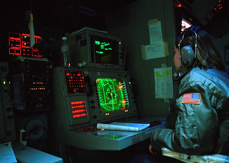
After the electrons pass through the sample they are expanded and hit a phosphor screen forming a green image. These phosphor screens might be more familiar to you from submarine movies that show green radar screens.
It is quite magical when you open up the column valve and see this green spot form on the saucer which is inside the high vacuum chamber. As you focus the image using a knob on the left of the console, an image of the molecules comes into focus. I have added in a Youtube video so you can also experience the excitement.
 |
| https://cemas.osu.edu/fei-tecnai-f20-stem |
Pictures are then taken on a camera hiding under the green screen which can be raised letting the electron beam focus onto a camera.
Making giant fullerenes
Below are the results for the heated sample. Collecting the mass spectrum for the heated soot using the mass spectrometer (which I spoke about in the last post) we found the distribution of mass peaks shifted towards higher masses. Imaging the edge of the soot using the electron microscope we could see small dark fringes which are the fullerene molecules. When you heat the sample, these small closed fringes are seen to enlarge. It is mind-blowing to think these cages are only a nanometre wide (that is 0.000000001 m, or a million times smaller than a metre). For some more help with the scale, you could line up 50,000 of these nanometre-sized cages and they would just be as thick as your hair.
We wanted to see the fullerenes as they were being heated to observe the tranformation in real time, so we increased the temperature of the wire in the electron gun, increasing the number of electrons boiling off the wire and flooding the sample with a very intense beam of electrons. This increased the temperature of the sample and allowed us to view the sample changing. You can see after 11 minutes of irradiation a smaller fullerene attached to a larger fullerene (indicated by the arrow) and then fused with it over the next nine minutes.
Here is one of the earlier videos I took of the computer screen as the sample was irradiated.
The observation of small fullerenes coalescing into larger fullerenes had never been observed before so it was very exciting to see this as it happened. The next blog post will involve some computer simulations to see how large, theoretically, these cages can become and also consider what we might do with them.


No comments:
Post a Comment